Fortunately, many types of cardiac diagnostic tests are available. Your clinician will order tests based on your specific medical situation. Tests include:
Blood tests
iStock.com/Artico
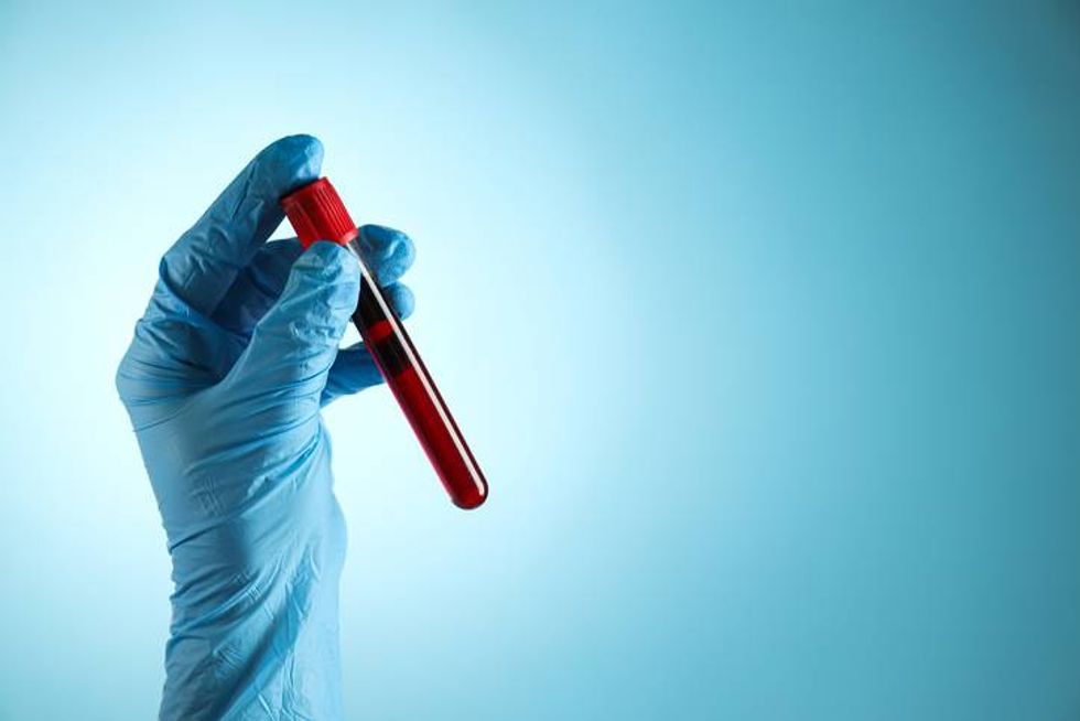
What it is: A needle inserted by a clinician into your vein to remove a small amount of blood.
What it measures: Cholesterol, proteins like lipoprotein (a) or troponin, and inflammation markers like C-reactive protein. Your clinician will order specific tests based on your possible diagnosis.
Used to: Assess your risk of cardiovascular disease, along with a physical exam and your medical history. Specific tests like troponin levels can confirm whether damage to your heart muscle has already occurred.
Blood pressure monitoring
iStock.com/bymuratdeniz
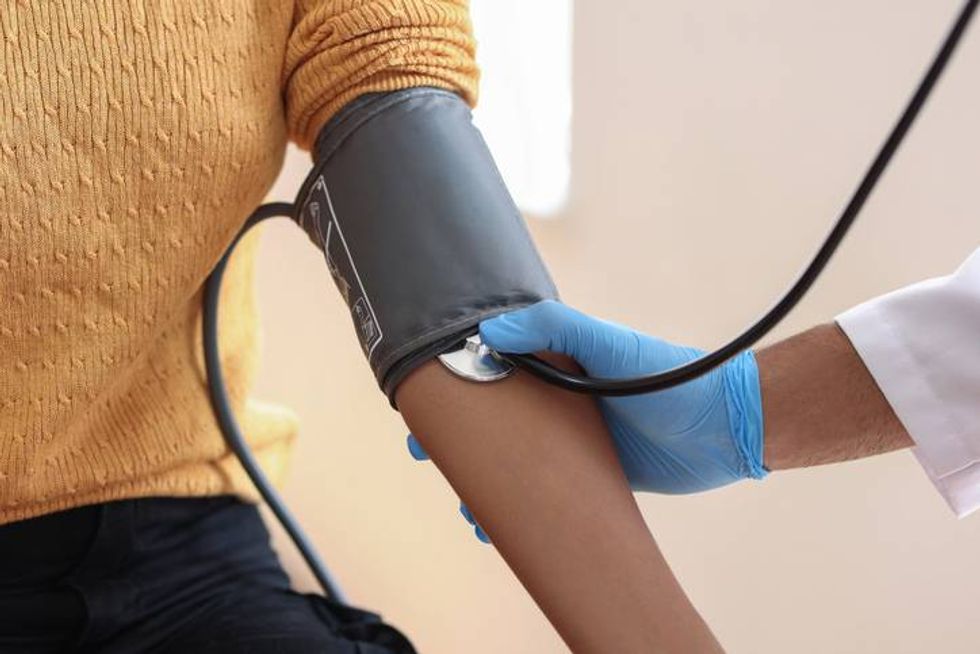
What it is: An arm cuff that measures your blood pressure at clinic visits. You may also need to take your blood pressure at home.
What it measures: How much pressure your blood puts on your arteries when your heart pumps.
Used to: Provide an early warning system of heart disease, even when there are no symptoms, since high blood pressure can damage your heart muscle and blood vessels over time.
Other uses: During a medical emergency, blood pressure monitoring can alert the team to sudden status changes.
According to the American Heart Association, blood pressure categories are:
Normal: Less than 120 mm Hg systolic (top number) and 80 mm Hg diastolic (bottom number) Elevated: 120–129 mm Hg systolic and less than 80 mm Hg diastolic Stage 1 hypertension: 130–139 mm Hg systolic or 80–89 mm Hg diastolic Stage 2 hypertension: 140 mm Hg or higher systolic or 90 mm Hg or higher diastolic Severe hypertension: 180 mm Hg or higher systolic or 120 mm Hg or higher diastolic
Chest X-ray
iStock.com/hut547
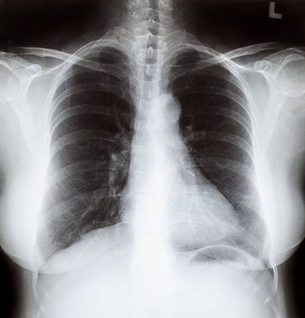
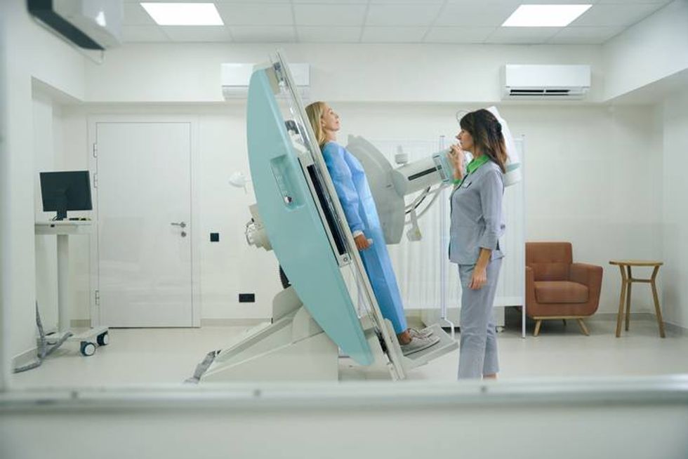
What it is: An X-ray that shows the heart, lungs and ribcage. It can’t show the structures inside the heart.
What it measures: A chest X-ray shows the size, shape, and location of the heart and lungs. Some nearby blood vessels will also appear in the image.
Used to: Help diagnose the cause of symptoms like chest pain, fever, breathing difficulties or a stubborn cough.
Echocardiogram (heart ultrasound)
iStock.com/Pitchayanan Kongkaew
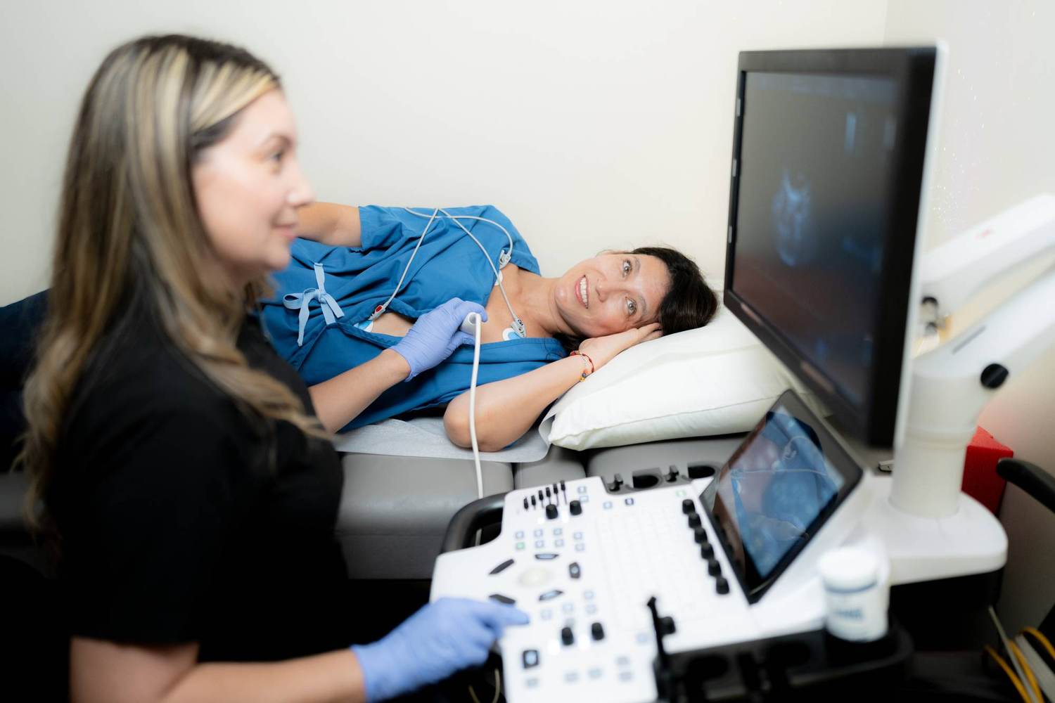
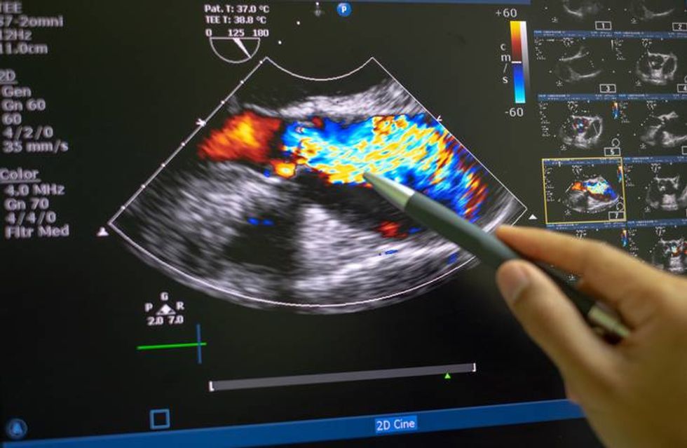
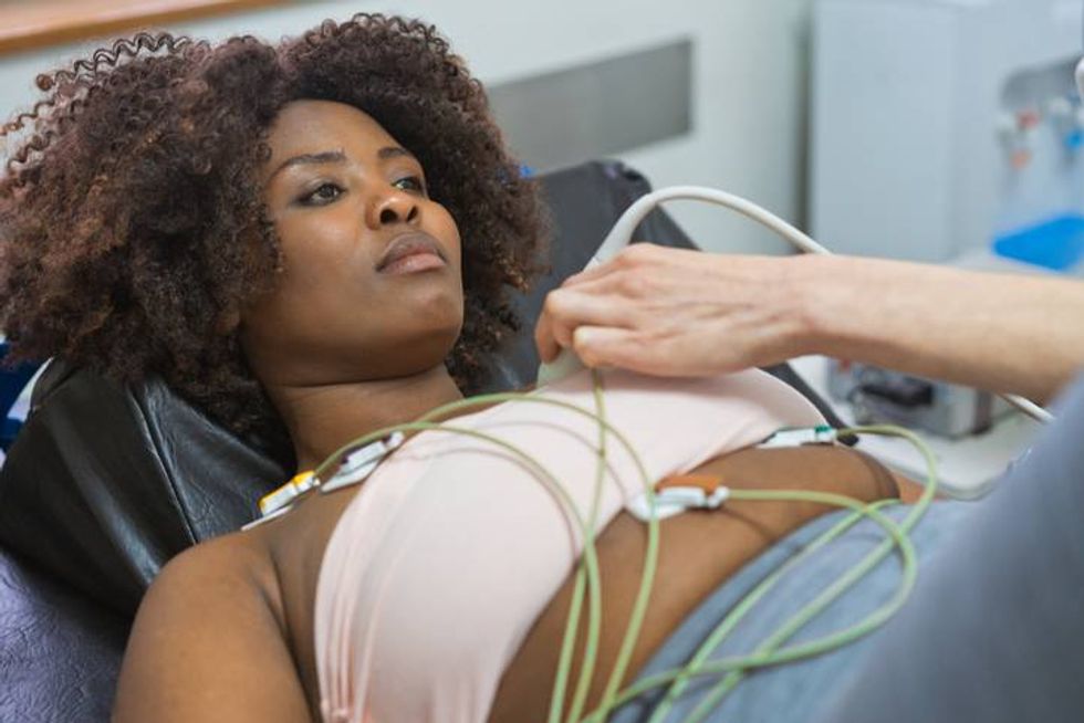
What it is: A test that uses sound waves to create pictures of the heart. The most common kind is called a transthoracic echocardiogram (TTE), where the clinician waves the ultrasound wand over your chest. There is also a transesophageal echocardiogram (TEE), where your clinician places the ultrasound wand down your throat to get a clear view of your heart or aorta.
What it measures: Your heart’s structure and how well it works, including its pumping strength and valve health.
Used to: Help diagnose heart muscle damage, heart valve disease or heart failure.
Electrocardiogram (ECG or EKG)
iStock.com/izusek
What it is: Electrodes applied to your body that are connected to an EKG machine.
What it measures: Electrical waves in the heart and how fast and regularly your heart beats. Each time your heart beats, it creates an electrical wave. This wave makes the heart muscle squeeze, which pumps blood from the heart.
Used to: Diagnose and monitor many heart conditions, including heart attack, heart failure, heart valve disease, coronary artery disease and heart arrhythmias (irregular heartbeats).
Other uses: Can help diagnose a pulmonary embolism, inflammation of the heart’s sac, or abnormal blood levels of potassium or calcium.
Tilt table test
iStock.com/yacobchuk
What it is: A test where you lie on a special motorized table that goes from flat to an angle that mimics sitting up. A technician will monitor your vital signs and symptoms like slow heart rate, low blood pressure, fainting, lightheadedness or nausea.
What it measures: Whether blood pooling in your veins causes these symptoms.
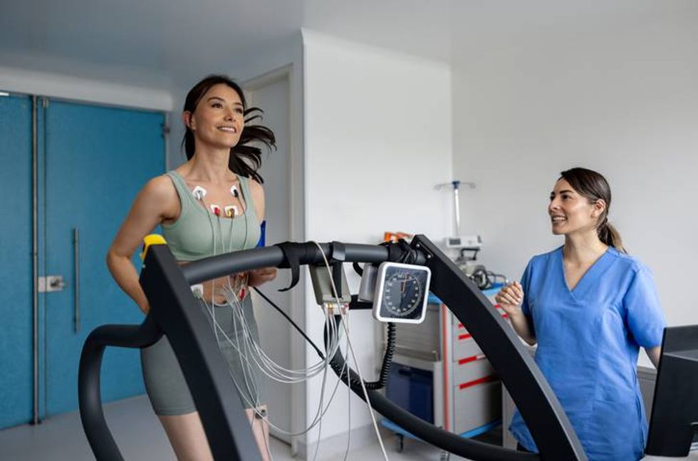
What it is: A test that requires walking on a treadmill or riding an exercise bike in a medical setting while hooked up to monitoring equipment. Your clinician will also check your blood pressure after the test.
What it measures: How well your heart pumps blood.
Used to: Diagnose coronary heart disease or find the cause of symptoms like chest pain or shortness of breath.
Other uses: Can also help predict the risk of a heart attack or eligibility for surgery or a safe level of exercise.
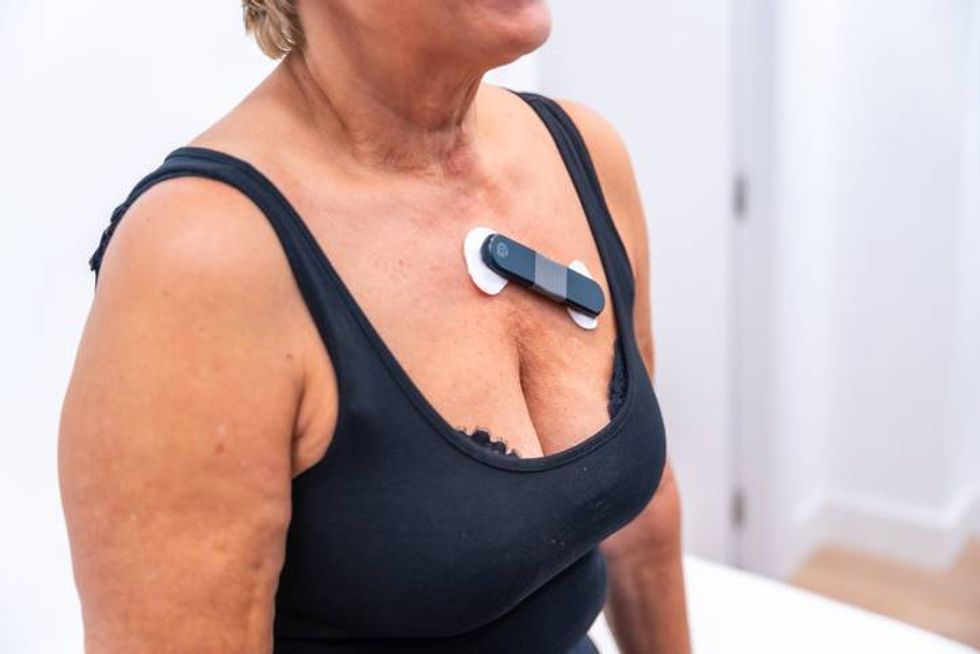
Holter monitoring
iStock.com/Unaihuiziphotography
What it is: A small wearable device that records your heart’s rhythm over 24 to 48 hours. Your clinician might order Holter monitoring if you have had fainting spells or dizziness, or an EKG did not provide enough information.
What it measures: Your heart’s rhythm throughout the testing period.
Used to: Detect irregular heartbeat (arrhythmia).
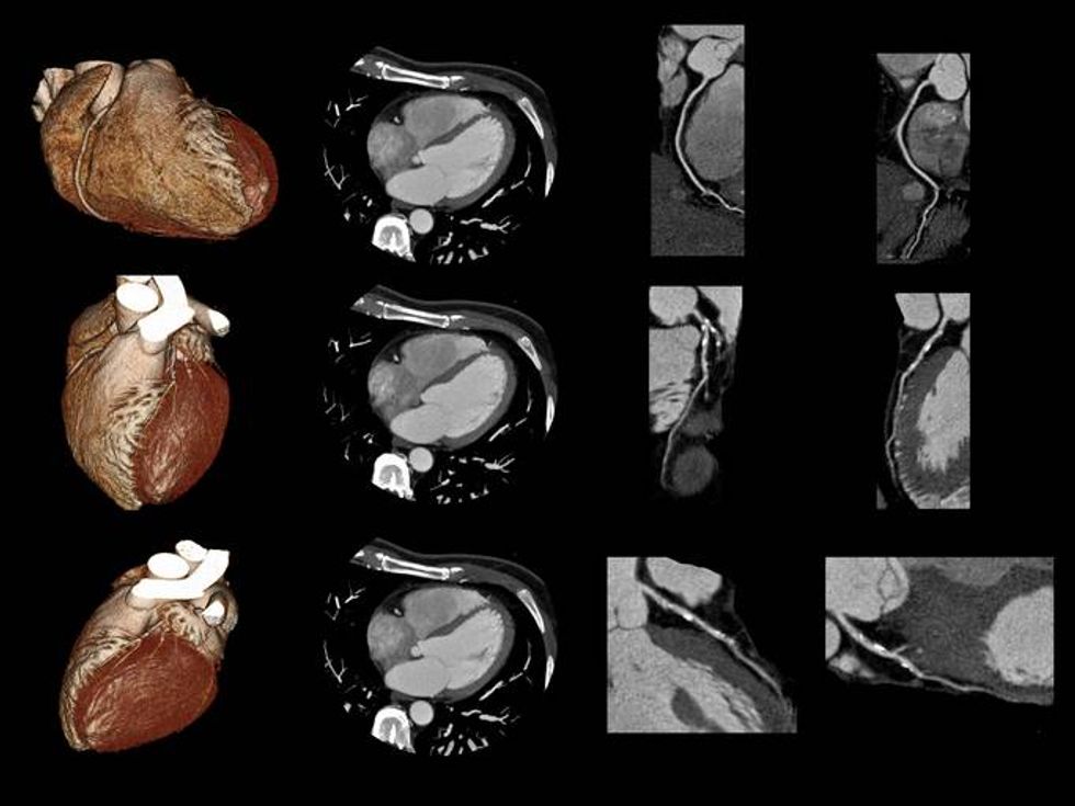
Cardiac CT scan
iStock.com/temet
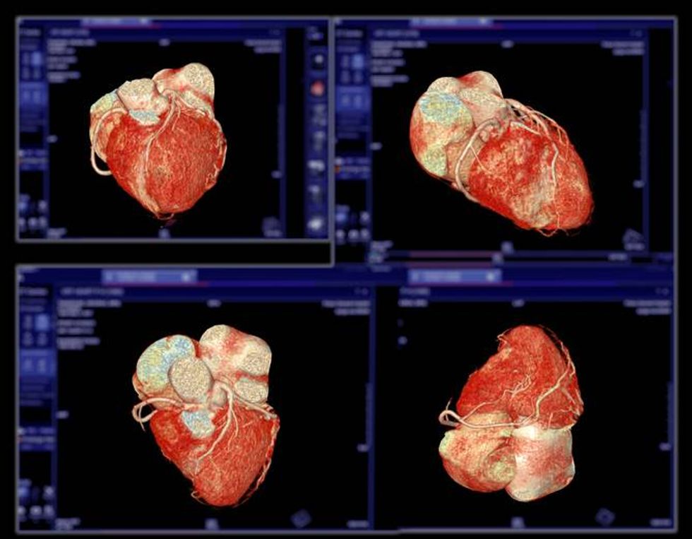
What it is: X-rays taken from many angles used to create a detailed 3D image of your heart and nearby blood vessels. Some high-resolution scanners can even show your heart in motion.
What it measures: The anatomy and function of your heart, valves, blood vessels and related structures.
Used to: Diagnose or monitor chest pain, blockages in your arteries, and problems with your aorta or heart valves.
Other uses: Often used to help plan cardiac procedures, including minimally invasive or robotic heart surgeries.
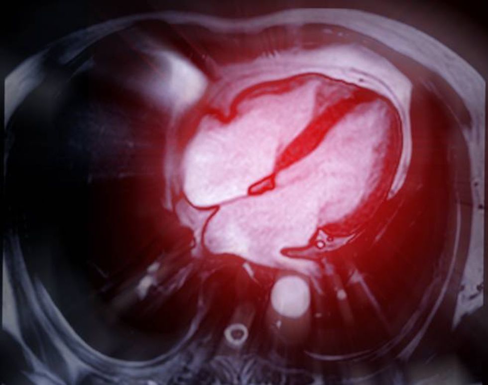
Cardiac MRI scan
iStock.com/mr.suphachai praserdumrongchai
What it is: A magnetic field plus radiofrequency waves used to create detailed 3D pictures of blood vessels and the inside of the heart, including valves.
What it measures: How well the heart and valves are functioning and whether enough blood is flowing through your major blood vessels.
Used to: Diagnose or monitor advanced or complicated heart conditions after other testing has been done.
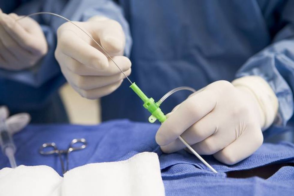
Cardiac catheterization
iStock.com/monkeybusinessimages
What it is: A small tube, or catheter, that gets inserted into one of your large veins to thread it up to your heart. Once there, the clinician will check your heart’s blood flow or the inside of your blood vessels for problems.
What it measures: How well your heart works and how healthy your valves and blood vessels are.
Used to: Diagnose blocked or narrowed arteries or valves.
Other uses: Unlike other diagnostic tests, cardiac catheterization allows your clinician to move from diagnosis to a corrective procedure immediately if needed. Possible procedures include clearing a blocked artery or partially blocked heart valve or taking tissue samples from the heart to study under a microscope.
Coronary angiogram
iStock.com/mr.suphachai praserdumrongchai
What it is: A specific type of cardiac catheterization. Once your clinician threads the catheter to your heart, they will take X-rays using contrast dye to check the large arteries near your heart for blockages.
What it measures: How healthy the arteries near your heart are.
Used to: Diagnose and then correct blocked arteries near your heart.
This educational resource was created with support from Merck.
Darcy Lewis
Darcy Lewis writes about health and medicine from Chicago, focusing on clinical medicine, the business of healthcare and health policy. A former National Cancer Reporting Fellow, she also completed the Medicine in the Media Fellowship at Dartmouth College in Summer 2024.
With a focus on helping patients make better healthcare decisions and helping clinical practitioners stay current on clinical developments, Darcy has gravitated toward writing about oncology. She has written about cancer for patient-facing publications like Everyday Health, Cancer Today and HealthyWomen.org, as well as professional-facing content and articles for ASCO Daily News, Oncology Live, Targeted Therapies in Oncology, Medscape, MedPage Today, McKesson, Cardinal Health, evi-Core and many others. One of her articles for Oncology Live won the 2020 ASJA award for best trade article, while an annual report for the Pulmonary Fibrosis Foundation won the 2024 ASJA award for long form content.
Darcy currently serves as ASJA’s president following stints as vice president, secretary and chair of the website task force, a multiyear effort to modernize ASJA’s communications with members and the public.