
Medical imaging is used by healthcare providers (HCPs) to view the body and gather information to diagnose and treat medical conditions. Understanding the different types of medical imaging can help you know what to expect.
X-ray
X-rays use radiation to quickly and painlessly create images of structures inside your body. They are commonly used to diagnose bone problems, breast cancer and infections.
- One type of X-ray called a bone density test (DEXA scan) measures the mineral content of your bones to see how strong they are. It is used to check for low bone mass (osteopenia) and weak, brittle bones (osteoporosis).
- Another type of X-ray, a mammogram, is breast imaging that’s used to detect breast cancer.
- Routine mammograms performed to check for breast cancer are called screening mammograms.
- Mammograms performed because a woman is showing signs of breast cancer, or after suspicious results on a screening mammogram, are called diagnostic mammograms.
What to expect:
Depending on the type of X-ray, you’ll be asked to lie down or stand still, and possibly switch positions, while the machine takes the images. You should be done in 10 to 15 minutes.
Tips and tricks:
- Wear loose clothing, and don’t wear anything with buckles, buttons or zippers. Metal can interfere with the test results.
- Don’t schedule a mammogram during the week before your period. It can be harder to get a good picture if your breasts are swollen, and your breasts may be more sensitive at this time.
- When going in for a mammogram, don’t wear deodorant, antiperspirant, powders, creams or perfume under your arms or around your breasts because they may show up as white spots on the X-ray.
Magnetic resonance imaging (MRI)
MRIs use magnetic waves to create detailed images of organs and tissue. They can be used to diagnose stroke and blood vessel issues, spinal cord problems, tumors, multiple sclerosis (MS), aneurysms, joint injuries, and more.
What to expect:
You’ll be asked to remove all your clothes and put on a hospital gown. Then, you’ll lie on a table that slides into a tunnel-like MRI machine, which may make loud noises as the magnets work. An MRI usually takes 45 minutes to an hour.
Tips and tricks:
- Talk to your HCP about options for helping you relax during your MRI, such as soothing music or a sleep mask.
- Some tests require you to be confined in a small space. Talk to your HCP about your options if you have any concerns about claustrophobia.
- You cannot have any metal in the room with you, since an MRI is a giant magnet. Be sure to alert the technician if you have any metal in your body.
PET scan
PET scans show how organs and tissues are functioning. Undergoing a PET scan involves inhaling, swallowing or getting a shot of radioactive medicine (called a tracer) so a machine can read the radiation these tracers give off. PET scans are often used to diagnose cancer, heart problems, Alzheimer’s disease, epilepsy and Parkinson’s disease.
What to expect:
After being given the tracer, you’ll lie on a table that slides into a donut-shaped scanner. The entire process will take around two hours.
Tips and tricks:
- You won’t be allowed to eat or drink for six hours before your PET scan (except plain water with nothing added).
- If you’re breastfeeding, you will not be able to breastfeed for 24 hours after getting your tracer because of the exposure to radiation.
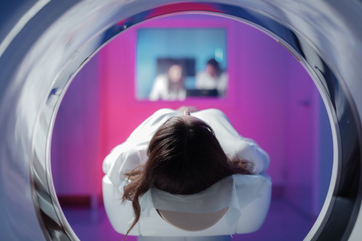
CT scan
Computerized tomography, or CT scans, use many X-ray images to create a cross-section of body parts, such as bones, blood vessels and soft tissues. CT scans are commonly used to assess traumatic injuries, fractures, tumors, infections, heart problems, cancer and more.
What to expect:
You’ll lie on a table that slides into a donut-shaped scanner, where an X-ray tube will rotate around you while taking images. CT scans are usually quick, lasting only about 10 to 15 minutes.
Tips and tricks:
- You can probably keep your own clothes on as long as you avoid wearing clothing with metal zippers or snaps. (Leave jewelry at home, including metal piercings.)
- Avoid eating and drinking for four hours before your CT scan.
Ultrasound
Ultrasounds use sound waves to capture images of structures in the body. In addition to helping monitor pregnancy, ultrasounds can be used to diagnose gallbladder disease, breast lumps, swollen joints, genital issues and more.
- One type of ultrasound, a transvaginal ultrasound, involves inserting the imaging device (called a transducer) into the vagina. This allows HCPs to see the organs inside your pelvic cavity, including your cervix, uterus, fallopian tubes and ovaries.
- During pregnancy, transvaginal ultrasounds are common during the first trimester (the first 12 weeks). After that, your HCP will likely want to do at least one abdominal ultrasound (also called a sonogram) to check your baby’s development.
What to expect:
An ultrasound technician will apply gel to your skin, then move the transducer around to create images of what’s going on inside your body. The ultrasound will probably last 30 minutes to one hour.
Tips and tricks:
- For some types of ultrasounds, you may be asked not to eat or drink before the procedure.
- For an ultrasound during pregnancy, you’ll likely need to show up to your appointment with a full bladder to help the technician get a better view of your baby.
If you have questions about healthcare imaging, talk to your HCP.