
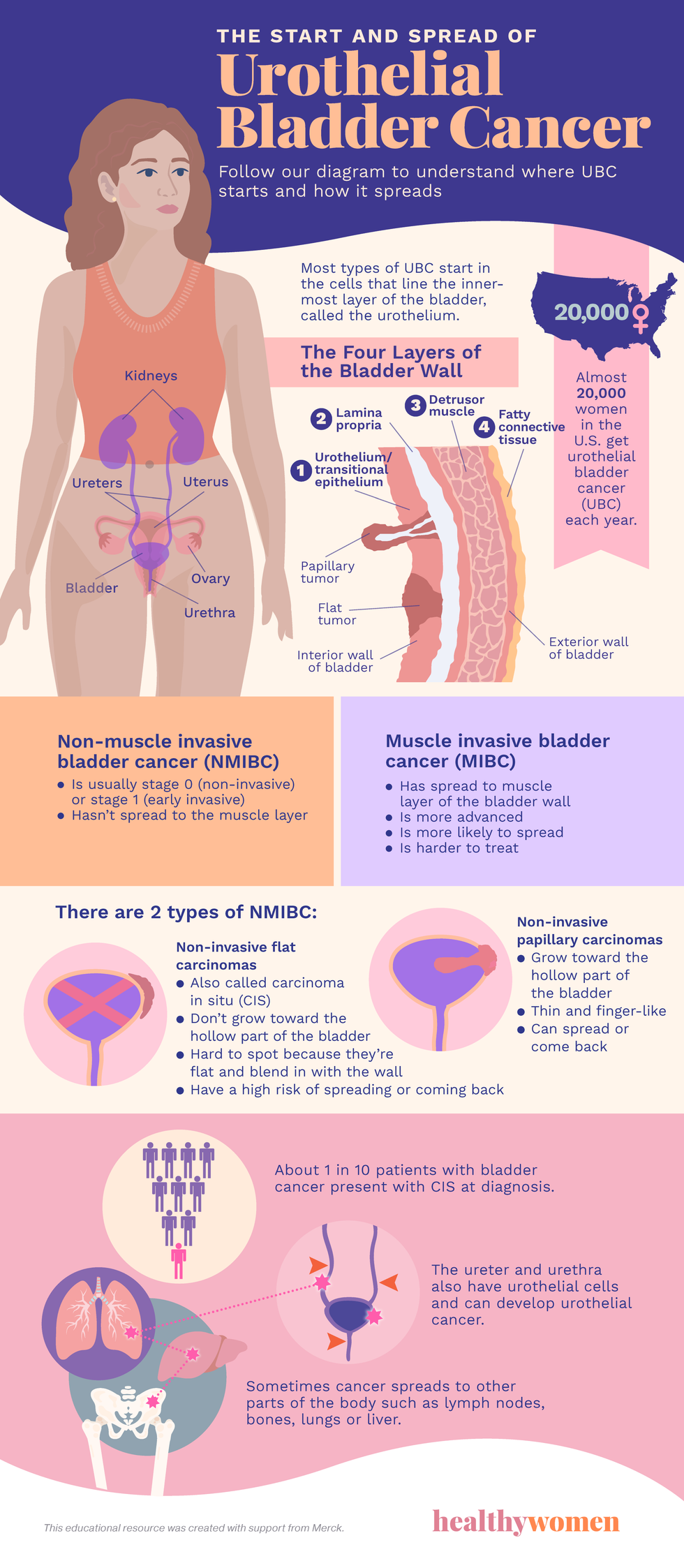
Almost 20,000 women in the U.S. get urothelial bladder cancer (UBC) each year.
The Four Layers Layers of the Bladder Wall
- Urothelial/Transitional epithelium
- Lamina propria
- Detrusor muscle
- Fatty connective tissues
Most types of UBC start in the cells that line the innermost layer of the bladder, called the urothelium.
Muscle invasive bladder cancer (MIBC)
- Has spread to muscle layer of the bladder wall
- Is more advanced
- Is more likely to spread
- Is harder to treat
Non-muscle invasive bladder cancer (NMIBC)
- Is usually stage 0 (non-invasive) or stage 1 (early invasive)
- Hasn’t spread to the muscle layer
There are 2 types of NMIBC:
Non-invasive flat carcinomas
- Also called carcinoma in situ (CIS)
- Don’t grow toward the hollow part of the bladder
- Hard to spot because they are falter and blend in with the wall
- High risk of spreading or coming back
Non-invasive papillary carcinomas
- Grow toward the hollow part of the bladder
- Thin and finger-like
- Can spread or come back
About 1 in 10 patients with bladder cancer present with CIS at diagnosis.
The ureter and urethra also have urothelial cells and can develop urothelial cancer.
Sometimes cancer spreads to other parts of the body such as lymph nodes, bones, lungs or liver.
This educational resource was created with support from Merck.
From Your Site Articles
- What Women Need to Know About Urothelial Bladder Cancer ›
- Life with Urothelial Bladder Cancer ›
- The Connection Between Smoking and Urothelial Bladder Cancer ›
- Fast Facts: Everything You Need to Know About Urothelial Bladder Cancer ›
- Clinically Speaking: Questions and Answers About Urothelial Bladder Cancer Treatment ›
- Immunotherapy for Bladder Cancer - HealthyWomen ›