In the United States, one in eight women will develop breast cancer in her lifetime. Diagnosis is closely tied to a woman's chance of survival, but diagnostic methods aren't always fast enough, precise enough or affordable enough for all. Lisa Poulikakos, 32, a postdoctoral materials scientist and engineer at Stanford University and a 2019 fellow in the L'Oreal USA For Women in Science fellowship program, is trying to solve this problem with nanotechnology and light.
Getting her start
Poulikakos was always fascinated by the physical world. But before exploring women's health through physical science, this fascination drew her to engineering and outreach work for women in STEM. When she arrived at the Swiss Federal Institute of Technology (ETH) in Zurich to start her undergraduate studies nearly 10 years ago, she was one of a handful of women in her engineering class.
To remedy this, Poulikakos started her university's first engineering group for women, which continues today. She organized networking events for her classmates and started an outreach program in which 80-100 female high school students from around the country come to ETH Zurich each year to learn about careers in engineering and their impacts.
For her own engineering career, Poulikakos decided to pursue mechanical and process engineering. "It's a very broad field with all kinds of applications [that] have to do with anything from developing the latest race car to nanorobots that can go into the human body," she said.
As she progressed in her studies, Poulikakos grew interested in how engineering might help solve health challenges. This line of thinking steered her work toward improving breast cancer diagnostics.
Poulikakos was also motivated by her grandmother's experience with breast cancer. "My grandmother's cancer was misdiagnosed because she did not have access to state-of-the-art technology," she said. By the time the doctors revised her grandmother's diagnosis, the cancer had advanced to an untreatable stage.
"That motivated me to use the power of engineering technology and nanotechnology to help patients like my grandmother access accurate diagnosis," she said. "Currently, breast cancer diagnostic technologies face trade-offs in precision, cost and time."
Bridging engineering and medicine
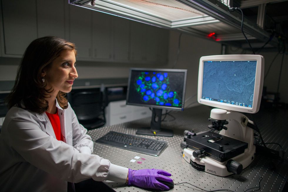
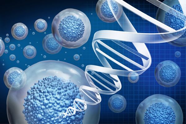
Poulikakos specializes in nanophotonics, which involves interactions between light and nanometer-sized objects. While conducting her Ph.D. research, also at ETH Zurich, she studied nano-optical materials. Nano-optical materials interact with light differently than materials without nanostructures on their surface, like plain glass, for example.
What caught Poulikakos' attention was how nanomaterials can scatter light in a way that allows scientists to detect the structural arrangement of molecules. While pouring over scientific papers on this subject, she found articles that discussed the optical properties of living tissues.
"That gave me the idea that we could also use nanoscale optical materials to address the challenges of tissue diagnostics," Poulikakos said.
Through the L'Oreal fellowship, Poulikakos has been bringing this idea to fruition. In order to diagnose many diseases, including breast cancer, doctors take a tissue sample, then stain or dye it to better see different cell structures under a microscope. A pathologist can arrive at a diagnosis within the hour at a low cost, but their assessment is subjective and not as precise as other techniques.
A different type of staining, biochemical staining, is more precise but costs more and takes longer. This method gives pathologists molecular and genetic information about the cancer in the tissue, which is important for determining a patient's treatment course. But it can take days for patients to receive their results, and patients who can't afford these expenses risk misdiagnosis with less precise methods.
Poulikakos wanted to develop a diagnostic technique that was not only compatible with current methods but also faster, more accurate and cheaper. She sought to replace the plain glass microscope slides pathologists use with nanopatterned ones. Similar to computer chip production, electron beams or light carefully etch the pattern into the slide surface.
Because of the surface's nano-optical properties, pathologists don't need to dye or stain tissue samples to see cellular details. When placed under a microscope, Poulikakos' surface scatters light through a tissue sample so that diseased and healthy tissue appear in different colors. "What the nanostructure does is similar to the way water droplets split light into a rainbow," she said.
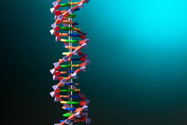
This interaction can show pathologists the arrangements of collagen fibers, a supportive protein found throughout the body, near tumors. This arrangement indicates whether a cancer is only affecting nearby tissue or if it has metastasized, or spread to other organs, and indicates whether a patient might respond to certain treatments.
"It would give a more accurate diagnosis of disease stage. The survival rate rapidly decreases for metastasized, advanced-stage disease," Poulikakos said. "That information would help inform the treatment in a more precise way."
When breast cancer is caught early, women have a 99% five-year relative survival rate. But if the cancer has spread to distant organs, that figure drops drastically — to 27%.
"The vision is to make accurate diagnosis more accessible," she said. "[Pathologists] can diagnose the disease more accurately and rapidly at a lower cost." Genetic and biochemical analysis would further add to the precision of Poulikakos' method.
Poulikakos and her team have started testing this diagnostic method using lab-made collagen that mimics human tissue. The next step is to test the technology on patient samples.
Her next steps
In November, Poulikakos will move to UC San Diego as an assistant professor, where she plans to build on this research. "I'm excited to start new projects which bridge engineering and medicine. I'm also excited to have the opportunity to work with the next generation of scientists."
Poulikakos credits her success to her mentors: her postdoctoral advisors. "I worked in teams led by two amazing women, leaders in both engineering and medicine," she said. "I hope to follow in their footsteps and be a role model for a more diverse, new generation of scientists."
She also plans to explore more ways to combine medicine and engineering. "There's great potential in bridging engineering and biomedicine," she said. "It's an exciting way to work toward a positive impact in health."