
True
Breast density is a term used to describe the amount of tissue versus fat in the breast. In dense breasts, there is more fibroglandular tissue — the lobules, ducts and connective tissue — and less fat. In general, women whose breasts have more than 50% fibroglandular tissue are said to have high-density or dense breasts.
Nearly half of all women aged 40+ who get mammograms have dense breasts. Women with dense breasts have a higher risk of breast cancer compared to women with less dense breast tissue, and the risk is higher with increasing breast density.
Breast density is not based on how your breasts feel and it has nothing to do with breast size or firmness. Dense breast tissue is seen only on a mammogram and appears as a white solid area that is hard to see through.
To determine the level of breast density, the radiologist who analyzes your mammogram will use the Breast Imaging Reporting and Data System (BI-RADS) to calculate the amount of dense tissue in your breast. BI-RADS classifies breast density into four groups:
A – Almost Entirely Fatty
This level means the breasts are almost completely made up of fatty tissue (less than 25% glandular tissue). An estimated 10% of women fall into this category.
B – Scattered Areas of Fibroglandular Density
Affecting about 40% of women, this level means there are scattered areas of density (from 25% to 50%), but most of the breast tissue is composed of fat.
C – Heterogeneously Dense
At this level, most of the breast tissue (between 51% and 75%) is dense. About 40% of women have this classification.
D – Extremely Dense
Affecting about 10% of women, this level indicates that more than 75% of the breast tissue is dense.
As you can see, it is common for women to have dense breasts. In fact, a large study published in the Journal of the National Cancer Institute estimates that more than 25 million U.S. women screened for breast cancer have dense breasts.
Who is likely to have dense breasts?
It is not clear why some women have a lot of dense breast tissue while others do not. What is known is that higher breast density is more likely if:
The link between high breast density and the risk of cancer
Breast density is a lesser known risk factor for breast cancer which should be considered in addition to other more widely known risk factors such as family history or genetic profile. Research has shown women with dense breasts are more likely to develop breast cancer compared to women with non-dense breasts, and the risk increases with increasing breast density.
Women, and especially women with dense breasts, should have a risk assessment conversation with their healthcare providers (HCPs), as there are validated risk assessment tools that take into consideration breast density. Individual risk assessment estimates a woman's 5-year, 10-year and lifetime risk of developing breast cancer and compares it to the general population risk. This is very helpful information. Women at elevated risk can take steps to lower their risk with lifestyle measures. Those who are at substantially increased risk may benefit from additional breast cancer screening and possibly even medication to lower their risk.
Today, three risk models are widely used. Designed by health professionals, these models take different approaches to understand a women's unique combination on risk factors as follows:
Using the information from one of these risk assessment tools, your HCP will be able to counsel you on your individual risk for breast cancer and make recommendations on when to start screening, how often to screen, and which screening tests to use.
For women with an elevated risk of developing breast cancer, mammography alone may not be adequate. Specifically, for women who are genetic mutation carriers, for those who have a strong family history of breast cancer or a history of prior chest radiation, and for those who have dense breasts and have a calculated lifetime risk (using risk assessment models) that is elevated (above 20% lifetime risk) mammography plus a second imaging method (MRI or ultrasound) may be appropriate. It is important for women to understand their risk, so they can discuss a more individualized approach to screening with their HCP.
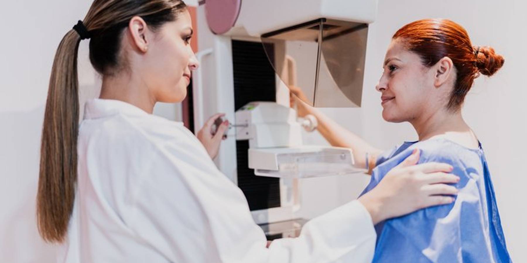
Mammograms
For women with an average risk of breast cancer, a yearly mammogram to screen for breast cancer is recommended. For women who have an average risk and dense breasts, supplemental imaging should be considered. Although most breast cancers can be seen on a mammogram, having dense breasts makes it harder for radiologists to see cancer on a mammogram. This is because dense tissue looks white on a mammogram, like a mass or tumor, making it difficult for the radiologist to tell the difference between a tumor and dense breast tissue. In contrast, fatty tissue looks almost black on a mammogram, so it is easier to see a tumor that looks white. Therefore, mammograms can be less accurate in women with dense breasts, producing false negative or false positive results, leading to either missed cancers or unnecessary follow-up tests.
According to several studies, three-dimensional (3D) mammograms are more helpful in finding breast cancers in dense breasts than the older two-dimensional (2D) mammogram. Compared to the 2D mammogram, where two X-rays are taken — one from the top and a second from the side — 3D mammograms collect multiple images of the breast from several angles, allowing doctors to see the breast tissue more clearly in three dimensions. As a result, breast centers are increasingly using 3D mammograms, especially in women with dense breasts. In fact, a recent study found that from 2015 to 2017, 3D mammography increased from 13% of screening examinations to 43%.
Supplemental Screenings
In general, screening mammograms miss one in five breast cancers in women and may miss one-third of breast cancers in women with dense breasts.
Therefore, many breasts specialists recommend adding supplemental screening tests along with mammography for women with dense breasts, based on evidence that the combination increases the detection of early breast cancer in women with dense breasts. These tests are mentioned below:
For women with dense breasts, the ASBrS recommends supplemental imaging such as breast MRI or ultrasound be considered in addition to annual mammography. For women with personal histories of breast cancer and dense breasts, the ACR recommends an annual breast MRI. Annual breast MRIs are also recommended for women with a known BRCA gene mutation, strong family history, prior chest radiation, or certain inherited disorders that increase the risk of developing breast cancer.
When considering possible screening options, the best strategy is to think in terms of the total breast cancer risk, including breast density. This means talking to your HCP about all your breast cancer risks so they can determine what tests are right for you. It is also important to find out if or when your health plan will cover supplemental imaging tests. While many states now require that women with dense breasts be covered by insurance for supplemental imaging tests, this is not universal.
Because having dense breasts is a risk factor for breast cancer, it is important to take charge of your breast health by learning about breast density and its implications for you. Ask about breast density at your next mammogram and make sure you get a copy of your mammography report, which will include an assessment of your breast density and categorize the amount of dense breast tissue in your breasts. Your HCP can also tell you if your mammogram shows you have dense breasts.
Also ask your HCP about the option of getting a risk assessment to determine your personal risk for breast cancer and the protective factors. The findings will help you and your physician weigh the pros and cons of additional screening tests. It is important to always remember to speak with your HCP about all your healthcare needs.
This resource was created with support from Bayer.
HealthyWomen content is for informational purposes only. Please consult your healthcare provider for medical advice, diagnosis or treatment.


Ductal carcinoma in situ – or DCIS – the earliest stage of breast cancer, is highly treatable. Here’s what you need to know.
El carcinoma ductal in situ o CDIS, la etapa más temprana de cáncer de mama, es altamente tratable. Aquí encontrarás lo que debes esperar durante el tratamiento y la recuperación.

Living with metastatic breast cancer has been hard, but I’m thankful for every day